History
In
1882 Robert Koch reported the discovery of the tubercle bacillus (4)
and described the appearance of the bacilli resulting from a complex
staining procedure. During the same time period several other
researchers (Ehrlich, Ziehl, Rindfleisch, and Neelsen), intending to
improve on Koch’s method, introduced modifications to the reagents and
the procedure. Franz Ziehl was the first to use carbolic acid (phenol)
as the mordant. Friedrich Neelsen kept Ziehl’s mordant, but changed the
primary stain to the basic fuchsin (first used by Ehrlich in 1882).
This method became known as the Ziehl-Neelsen method in the early to mid
1890s. In this method heat is used to help drive the primary stain
into the waxy cell walls of these difficult-to-stain cells. The use of
heat in this method has been the reason that this technique is called
the “hot staining” method.
The Ziehl-Neelsen method has endured as a reliable and effective way to demonstrate the acid-fast bacteria.
In
1915, Kinyoun published a method that has become known as the “cold
staining” method because the heating step was removed in favor of using a
higher concentration of the carbolfuchsin primary stain.
Purpose
The
acid-fast stain is performed on samples to demonstrate the
characteristic of acid fastness in certain bacteria and the cysts of Cryptosporidium and Isospora. Clinically, the most important application is to detect Mycobacterium tuberculosis in sputum samples to confirm or rule out a diagnosis of tuberculosis in patients.
Theory
There
are three common acid-fast staining methods, Ziehl-Neelsen (hot),
Kinyoun (cold), and Auramine-Rhodamine Fluorochrome (Truant method).
The emphasis in this Atlas-Protocol project will be on the Ziehl-Neelsen
and the Kinyoun methods because the slides produced by these methods
can be visualized using a standard bright-field microscope. The
fluorochrome method is used by large laboratories that have a
fluorescent (ultraviolet) microscope. For comparison purposes, the
recipe for the reagents and the protocol for all three methods are
included below, but images for the fluorochrome method will not be a
part of the Atlas at this time.
Many
bacterial cells are easily stained with simple stains or using the Gram
stain. A few types of bacteria, such as the mycobacteria and Nocardia
species, do not stain using these techniques or, if stained, they
produce a variable reaction because their walls are not permeable to the
rosaniline dyes in common staining regimens (12). The cell walls of
the mycobacteria contain mycolic acids giving the cell walls a high
lipid content. This characteristic is thought to be the reason (5, 10)
these bacteria are difficult to stain. To view these cells in samples
staining requires higher concentrations of the dye solution and/or a
heating period (4). However, once a stain is introduced into the cell
wall, removing it with a decolorizer is even more difficult. The
expression “acid fast” is derived from the observation that even with
the addition of hydrochloric acid to the alcohol decolorizer, some of
the stained cells retain the primary stain (carbolfuchsin). Cells that
release the primary stain (carbolfuchsin) with decolorizing will be
visible after the counterstaining step is complete. Bacteria described
as acid fast will appear red when examining specimens using bright-field
microscopy. Non-acid-fast cells and field debris will appear blue.
Acid
fastness is a characteristic that is shared by just a few organisms, so
staining to determine if organisms possess this trait is useful in
microbial identification schemes.
Acid-fast Staining
 Introduction
by: Marise A. Hussey and Anne Zayaitz |
Procedure
The Acid-fast Stain:
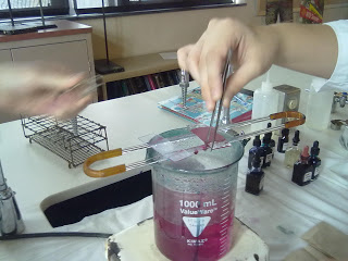
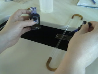
To start this procedure, we once again began with the steps for making a simple stain. After, we placed the slide over a beaker of boiling water suspended by a slide drying rack. Then we covered the slide with Bibulous paper and saturated the paper with Ziehl-Neelsen Carbolfuchsin.
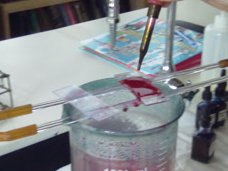
Like the last procedure, we had to keep the slide saturated with the dye while it steamed, but this time for 3-5 minutes. After the allotted time we removed the slide carefully from the rack and removed the Bibulous paper, throwing the used paper away in the designated receptacle.
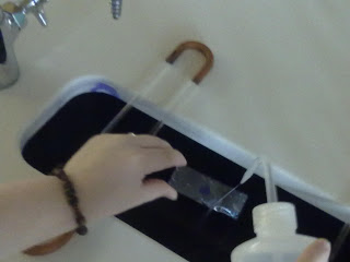
After cooling, we rinsed the slide with deoxidized water to remove the excess stain. Also to remove more stain, we used Acid-alcohol. To do this we held the slide at an angle over the sink, and added Acid-alcohol to it until the magenta color stopped running.
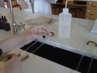
Immediately we rinsed the slide free of decolorizing agent so that the rest of our process was not inhibited. We then covered the slide directly with Methylene Blue for 2 minutes. After this time frame, we rinsed the slide clean of excess dye and blotted the slide dry.
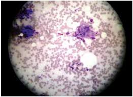
The Resluts:
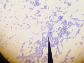
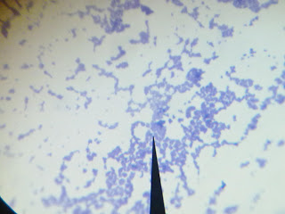
Once again the small cocci colonies are present within our slide, but this time with a different stain color and meaning. The blue appearance reveals that our bacteria is Non-acid-fast. If the magenta stain remained after using the Acid-alcohol, then our bacteria would be considered Acid-fast. In an Acid-fast bacteria the first dye (red) is trapped by waxes in the cell membrane.
The Acid-fast Stain:
To start this procedure, we once again began with the steps for making a simple stain. After, we placed the slide over a beaker of boiling water suspended by a slide drying rack. Then we covered the slide with Bibulous paper and saturated the paper with Ziehl-Neelsen Carbolfuchsin.
Like the last procedure, we had to keep the slide saturated with the dye while it steamed, but this time for 3-5 minutes. After the allotted time we removed the slide carefully from the rack and removed the Bibulous paper, throwing the used paper away in the designated receptacle.
After cooling, we rinsed the slide with deoxidized water to remove the excess stain. Also to remove more stain, we used Acid-alcohol. To do this we held the slide at an angle over the sink, and added Acid-alcohol to it until the magenta color stopped running.
Immediately we rinsed the slide free of decolorizing agent so that the rest of our process was not inhibited. We then covered the slide directly with Methylene Blue for 2 minutes. After this time frame, we rinsed the slide clean of excess dye and blotted the slide dry.
The Resluts:
Once again the small cocci colonies are present within our slide, but this time with a different stain color and meaning. The blue appearance reveals that our bacteria is Non-acid-fast. If the magenta stain remained after using the Acid-alcohol, then our bacteria would be considered Acid-fast. In an Acid-fast bacteria the first dye (red) is trapped by waxes in the cell membrane.
References:
http://www.microbelibrary.org/component/resource/laboratory-test/2870-acid-fast-stain-protocols
http://samsarahmicrobio.blogspot.com/2011/10/creating-endospore-and-acid-fast-stain.html
http://www.microbelibrary.org/component/resource/laboratory-test/2870-acid-fast-stain-protocols
http://samsarahmicrobio.blogspot.com/2011/10/creating-endospore-and-acid-fast-stain.html